Chrysotile Asbestos
Ted M. Clarke
![]()
![]() Double Click on Images for larger images.
Double Click on Images for larger images.
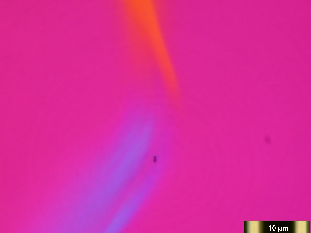
Here are some images of Chrysotile
asbestos in 1.550HD mountant that show use of this technology for mineral
identification.
"The plane polarized darkfield image shows the
characteristic dispersion staining color and this method works near the limit of
the resolving power of the microscope. These images have not been
published but might be in the future. Optical dispersion staining at this
resolution has never been done before. These images of the same field were
taken using the LOMO 60 X 1.00 NA oil immersion apochromat using my modified
LOMO Biolam. My punched polymer film analyzer and red wave plate work very
well, are cheap and easy to make.They also precisely orient in the plastic sliders."
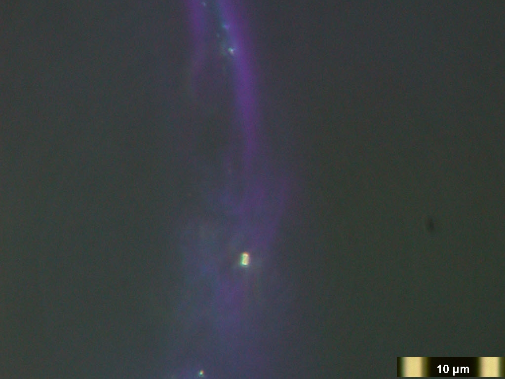
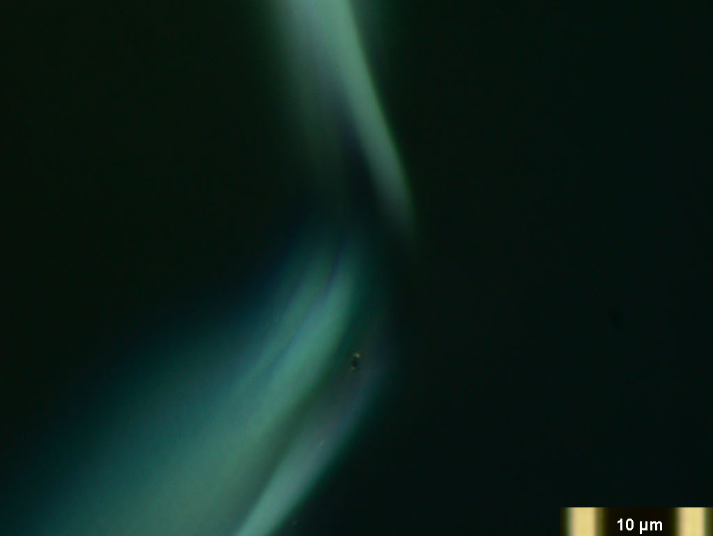
A darkfield image of a Chrysotile asbestos specimen in 1.550HD
index mountant.
"You can see that the oil immersion 1.25 NA
objective can be used at full resolution without a funnel stop using
darkfield above 1.25 NA provided by the LOMO 1.40 NA condenser with the
right size stop. I see no need for me to own a darkfield
condenser, especially when none of them have a high enough NA to use
with a 1.25 NA objective without a funnel stop to reduce the NA to 1.0.
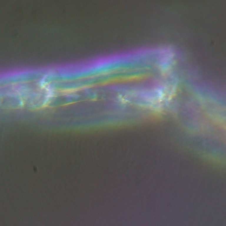
The digital images are intended to show the subjects as they appear through the
eyepieces. The default settings of the Nikon CoolPix 995 digital camera
are used without subsequent enhancement, although adjusting contrast and
reducing brightness can improve the darkfield images."